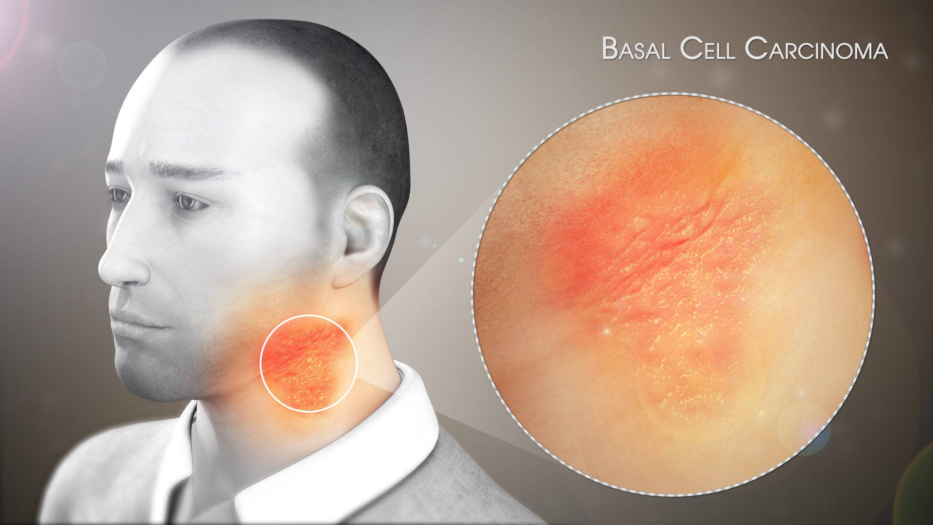
Basal Cell Carcinoma or BCC is a kind of skin cancer that originates in the basal cells -- the type of cells that line the epidermis and perform the routine function of replacing the old cells with the new ones. In this medical condition, the cancer of the basal cells appears as tumors on the skin’s surface. These tumors resemble sores, growths, scars, red peaches, or bumps. Basal cell carcinoma is the most widespread form of skin cancer in humans.

Symptoms
BCC is known to develop on those parts of the human body that are frequently exposed to the sunlight, i.e. ultraviolet (UV) rays. The areas on which it is more likely to develop are the facial region, shoulder, neck, arms, scalp, or ears. Sometimes BCC emerges in those areas that are not exposed to sunlight.
The only major symptom of BCC is growth on the skin or change in its appearance. These growths are generally painless. There are different forms of BCC and the symptoms of each form are peculiar and different, as mentioned below:
- Pigmented BCC: In this type of BCC, black, brown, or blue lesions appear on the skin. These lesions often have a translucent and raised border.
- Nonulcerative BCC: In this type of BCC, white, pink, or skin-colored bulges appear on the skin. Sometimes, the bulges are translucent and blood vessels beneath are visible. This type of BCC is very common and appears on the ear, face, and neck. Sometimes, these can burst, bleed, and scab over.
- Morpheaform BCC: It is the least common form of BCC in which scar-like lesions appear on the skin with a waxy and white-colored appearance with no border. This type of carcinoma can indicate an invasive form of BCC, which most probably can be disfiguring.
- Superficial BCC: People with this type of BCC have reddish patches on their skin, which are scaly and flat, and grow continuously. The patches often have a raised edge which usually emerges on the back or the chest.
- Basosquamous BCC: This type of carcinoma has the characteristics of both BCC and squamous cell carcinoma -- another type of skin cancer. It is very rare but is more likely to spread to other body parts when compared with other types of skin cancer.
Causes
Basal cell carcinoma, like almost every skin cancer, is generally caused due to long-term exposure to sunlight or ultraviolet rays exposure. Sometimes, intense occasional exposure resulting in sunburn can also lead to the development of the disease. People affected with BCC have the highest probability of recurrence.
Some other causes of BCC may include:
- Radiation exposure
- Skin conditions with chronic inflammation
- Arsenic exposure
- Complications from scars, infections, tattoos, burns, and vaccination
One of the major reasons behind BCC is a mutation in the DNA of the basal cell, where it will multiply rapidly and continue to grow when it would normally die. Eventually, these abnormal cells form a cancerous tumor, that appears as a lesion on the skin.
Treatment
As per several medical resources, doctors and physicians treat basal cell carcinoma by getting rid of the growth. Treatment of the disease depends upon the type of BCC, size, and location of the lesion, which includes:
- Curettage and electrodesiccation (C and E): In this method, the growth is scraped off with a curette (scraping instrument) and burned with an electrocautery needle, leaving a round white scar. This treatment is very effective especially on small lesions that are less likely to recur.
- Cryosurgery: In this method, doctors freeze and kill cancerous cells in the affected area with liquid nitrogen. This treatment is mostly prescribed for cancers that are thin and don’t extend far into the skin, and for people with bleeding disorders. This has a risk of loss of sensation in the area.
- Excisional surgery: In this procedure, doctors remove the tumor and its surrounding border of normal skin with a scalpel. Stitches are made in the area to close the surgical site, which may leave a little scar. It is often used for more advanced forms, which are at risk for affecting the surrounding skin.
- Mohs micrographic surgery: In this procedure, the tumor is removed layer by layer. Doctors take out some tissue, then look at it under a microscope to see if it has cancer cells, before moving on to the next layer. It is mostly used for large tumors or tumors in highly visible areas like the face or neck.
Disclaimer: The information in no way constitutes, or should be construed as medical advice. Nor is the above article an endorsement of any research findings discussed in the article an endorsement for any of the source publications.


