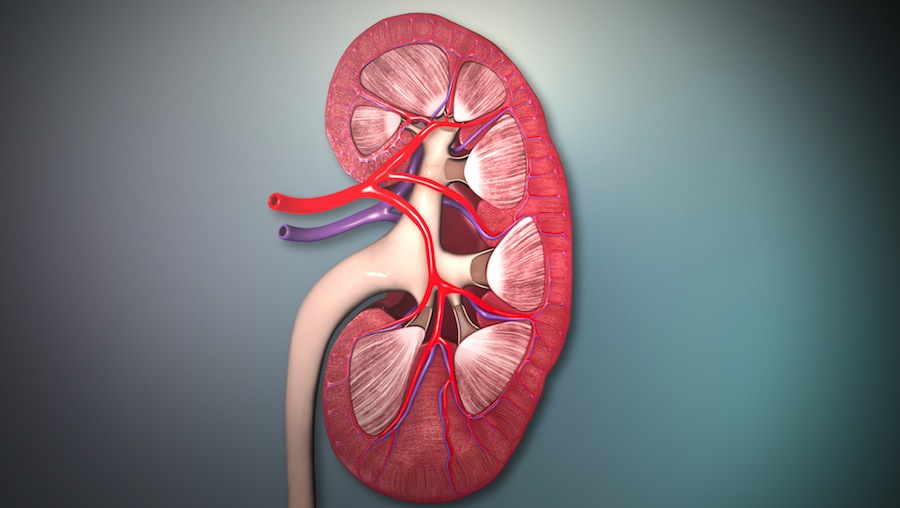
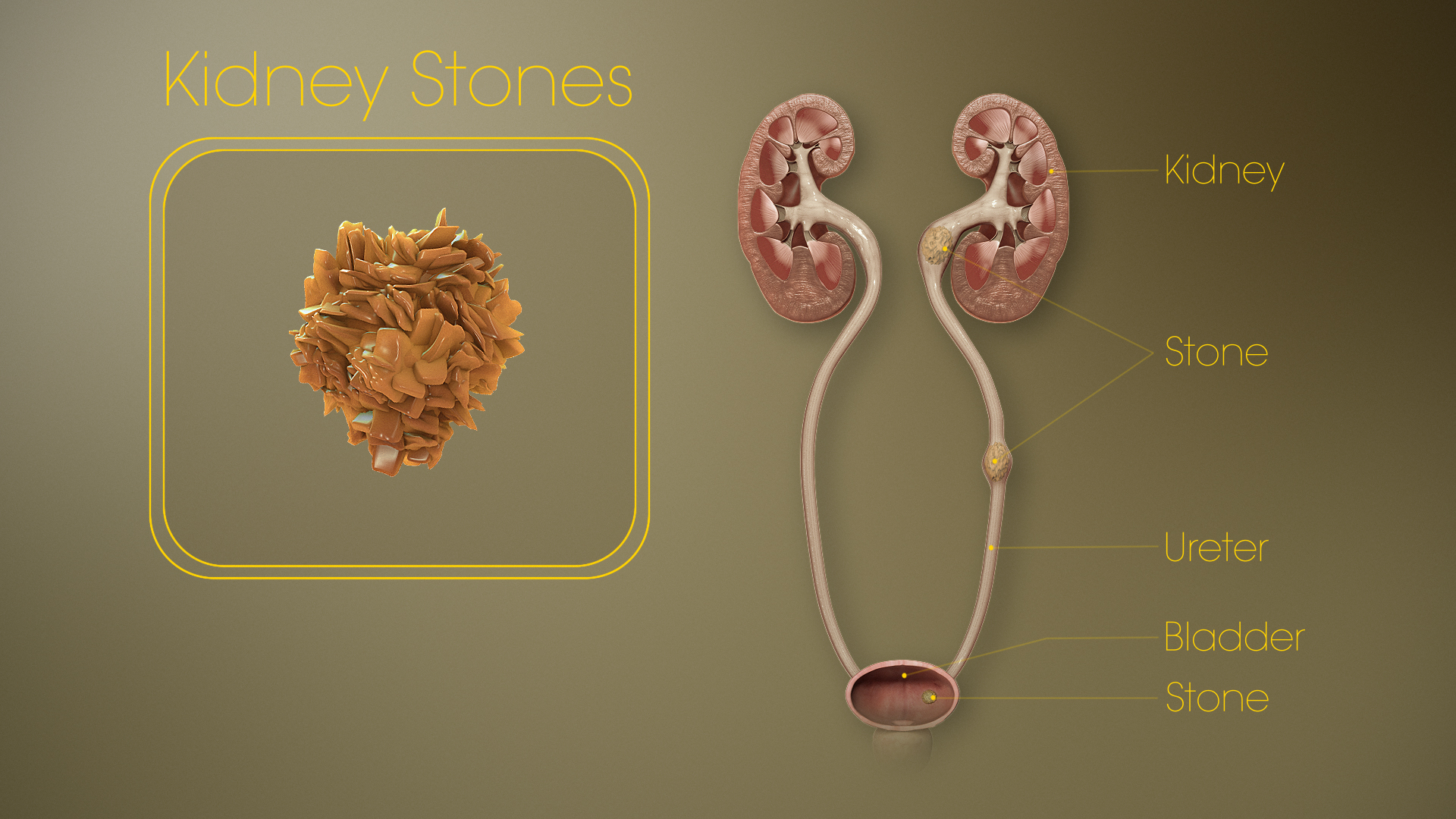
Kidney Stones (Scientific Name: Renal Calculus) results from an amassing of dissolved minerals that accumulate on the inner surface of kidneys. Depending on the amount of deposit, these stones can grow as big as a golf ball. They have a sharp and crystalline structure, and generally comprise of the compound calcium oxalate. The stones can pass through urinary tracts but may often cause unbearable pain. If they remain inside the kidney, these stones need to be operated out of the body in order to clear the ureter and allow for the passage of urine.

3D Medical Illustration – Cross section of the kidney
Causes of Formation
Kidney stones are formed if variations occur in the standard balance of water, salts and minerals in the body. Any imbalance in a person’s metabolism can cause an abnormal rise of mineral salts in the urine. Depending on the change in balance, kidney stones are formed. The most common kidney stone type is the calcium-type, which is formed due to calcium imbalances in the urine.
The factors that cause kidney stones are:
- Irregular intake of water. Due to low levels of water in the body, the salts, minerals and other substances form a mass, which gradually develops into a stone. Regular intake of water is thus very important.
- Various medical conditions are also often responsible for kidney stones. The uric acid stone type, for example, is caused in people with diseases like gout, inflammatory bowel diseases like Crohn’s disease, or some form of cancer.
- People suffering from hyperparathyroidism, in which the parathyroid gland (which secretes calcium regulating hormone PTH in the body) remains overactive, are also susceptible to kidney stone formation
- Kidney stones can also be a hereditary problem.
Diagnosing Kidney Stones
People facing intolerable pain while urinating should check for the symptoms of kidney stones.
Symptoms of kidney stones include:
- Spasms of intolerable pain in the back below the ribs and around the abdomen, reaching out to the groin or genitals
- Persistent aching at the sides of the back
- Passage of blood in urine
- Urinating in unusually short intervals and severe pain during urination
- Nausea
A doctor will diagnose and check whether the pain is caused by kidney stones or some other health problems are at fault.
The procedure for diagnosing kidney stones involves a few basic tests, such as:
- Blood tests that will ensure whether the kidneys are functioning well, and check the level of minerals and other substances that could cause kidney stones
- Urine tests that will check for any kind of urinary infections, or the presence of ‘invisible’ stones in the urine
- Diagnosis of the stones that flow out with the urine, and examination of the stone-types to decide on the kind of treatment that the patient needs
A few other tests could be involved to confirm the diagnosis and situate the kidney stones in the body, which include X-ray, an ultrasound scan, a Computed Tomography or CT scan, or an Intravenous Urogram (IVU). IVUs and CT scans are presently the most accurate techniques of locating blockages.
Treatments for kidney stones
The treatment for kidney stones depends primarily on the type and size of these stones. If the stones are small, a person can easily pass these stones through urine. The smaller stones cause pain which lasts for a day or two and then subsides as the stones flow out.
People facing terrible pain are injected with painkillers and anti-emetics to treat the nausea. If the stones are small in size and do not need to be surgically removed, the doctor may advise waiting until it passes with the urine.
It is also advisable to drink a lot of water to avoid further growth of stones. Yellowish or brownish urine signals insufficient intake of water.
Kidney stones that are larger than 6-7mm, need to be removed surgically. There are various techniques to remove the larger stones, depending on the size and location of these stones in the body.
The different kinds of treatments are:
- Extracorporeal shock wave lithotripsy (ESWL): Using X-rays or ultrasound, the kidney stone is located, and then shock waves of energy are sent to the stone to crush it into tiny pieces. This facilitates the passage of the stone through urine. It is often uncomfortable and the patient may need painkillers during the treatment. More than one session is required to remove the stone completely.
Success rate: ESWL is 99% effective on 20mm diametric stones. - Ureterorenoscopy or retrograde intrarenal surgery (RIRS): This treatment is for stones that are stuck in the ureter. The treatment involves the passing of a long, thin telescope (ureteroscope) through the urethra, into the bladder and then on into the ureter from where the stone has to be removed. With the help of another small instrument or laser energy, the stone can be removed. Anesthesia is used in this treatment to ease the patient’s pain.
Success rate: RIRS is 50-80% effective on stones up to 15mm.
3D Medical Animation – Kidney stone treatment
- Percutaneous nephrolithotomy (PCNL): This treatment is used for larger stones when ESWL seems unsuitable for the patient. An incision is made on the back to lead to the kidney. Using a nephroscope (a thin telescopic instrument) the stone is reached through the cut, and like in RIRS the stone is removed with an instrument or crushed with laser energy. The patient is given anesthesia in this case as well.
Success rate: 86% effective for stones up to 21-30-mm.
- Open surgery: Open surgery is required in very rare cases, when the stone is unusually large or the patient has some anatomical abnormality. An incision in the back leading to both ureter and the kidney is made, and then the stone is surgically removed.
There is a probability of having kidney stones again, after getting them removed. To avoid the recurrence:
- Continue medication with doctor’s advice.
- Avoid dehydration, excess salt intake, drinks with phosphoric acids, tea or coffee, mineral water.
- Drink citrus juices regularly.


